 Türkçe
 English

Blog
Bursa Impacted Tooth Extraction: Surgical Operation and Clinical Processes
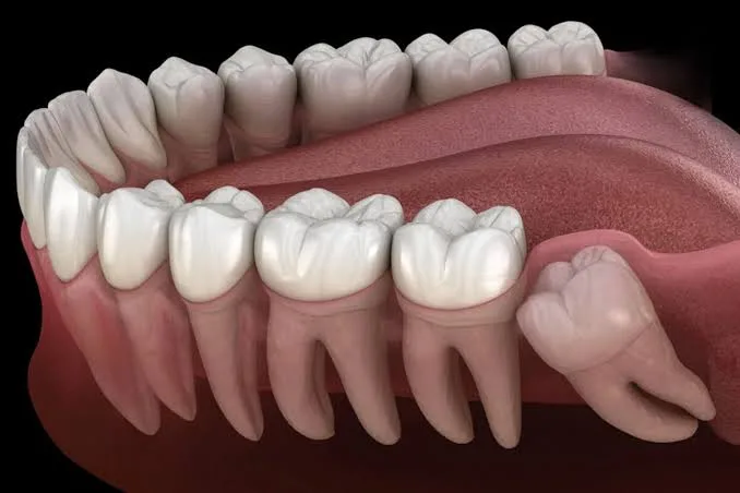
The development of the jawbones and the eruption process of teeth (their emergence into the mouth) in human anatomy are directly influenced by genetic factors and evolutionary changes. A lack of sufficient space in the jawbone, the early or late loss of primary teeth, or anatomical deviations in the eruption axis of a tooth can cause some teeth to fail to reach the oral cavity and become trapped under bone or gum tissue. In dental terminology, this condition is called an "impacted tooth".
If you are experiencing unexplained pain in the jaw area, recurrent gum swelling, or orthodontic problems and are looking for an objective, scientific source about the surgical processes of Bursa impacted tooth extraction, this guide covers the anatomical foundation of the operation and the treatment stages in full detail.
This article has been prepared in full compliance with health services information regulations and medical ethics rules, with the aim of informing the public about oral, dental, and maxillofacial surgery applications.
Which Teeth Become Impacted and Why?
When people think of impacted teeth, wisdom teeth (third molars) usually come to mind first. Because the evolutionary reduction in the size of our jaw structure has left no room for these last erupting teeth, wisdom teeth are the group most commonly impacted. However, contrary to popular belief, the risk of impaction is not limited to these teeth alone:
Canines: Located at the corners of the jawbone and serving as key elements of the chewing system, canines are the second most commonly impacted tooth group after wisdom teeth. They are usually trapped within the bone on the palatal or labial side in the upper jaw.
Premolars: They may become impacted, especially as a result of primary teeth being extracted too early, causing neighboring teeth to drift and close off the space for the permanent tooth coming in underneath.
Supernumerary Teeth: These are extra teeth that develop beyond the number normally present in human genetics (32), usually forming within the jawbone between the front incisors (mesiodens).
When Should Impacted Teeth Be Removed?
Some teeth that are completely impacted within the bone and do not harm surrounding tissues (adjacent tooth roots, nerves, or sinus cavities) and show no pathological signs (such as a cyst) may be left in the mouth under a physician's supervision and monitored with periodic X-rays. However, in most cases, surgical extraction becomes a medical necessity. The main situations that indicate extraction are as follows:
Cyst and Tumor Formation: The protective sac surrounding the impacted tooth, called the follicle, may gradually fill with fluid and turn into cystic lesions. These cysts can grow silently, leading to resorption in healthy jawbone and creating a risk of jaw fractures.
Resorption of Adjacent Tooth Roots: An impacted tooth attempting to erupt in a horizontal or angled position exerts constant physical pressure on the root of the healthy tooth in front of it. This pressure causes irreversible root resorption and severe pain in the healthy tooth.
Infection and Pericoronitis: In cases where only a small part of the tooth has broken through the gum (semi-impacted), significant bacterial buildup begins in that area. Food debris accumulating in the gum pocket can lead to acute inflammation called "pericoronitis," facial swelling, and jaw locking (trismus).
Need for Orthodontic Treatment: In patients who will undergo braces or clear aligner treatment, problem-causing impacted teeth need to be surgically removed in order to create space in the jaw for the teeth to move. In addition, upper impacted canines can in some cases be brought into eruption and saved with orthodontic forces (pulled into the mouth), while those at an angle that cannot be saved are removed surgically.
Degrees of Impaction (Retention Types)
The most important factor determining the difficulty of the operation and the clinical planning is the tooth's position beneath the tissues. Impaction is divided into three main categories:
Soft Tissue Retention: The tooth has completely passed through the jawbone, but it is covered by a thick layer of gum tissue. No bone removal is needed; the tooth is exposed and extracted with only a small gum incision.
Partial Bone Retention (Semi-Impacted): Part of the tooth is visible in the oral environment, while part of it remains inside the jawbone. The bone tissue blocking the tooth's path must be removed with small burs.
Complete Bone Retention: The tooth has never reached the oral cavity and remains entirely deep within the jawbone. Removing these teeth requires advanced maxillofacial surgery protocols.
Bursa Impacted Tooth Extraction: Clinical Procedure Steps
Impacted tooth surgery involves biomechanical principles that are completely different from a standard tooth extraction. The clinical steps applied in maxillofacial surgery centers in Bursa are as follows:
1. Three-Dimensional Radiographic Diagnosis (CBCT)
Traditional two-dimensional (panoramic) X-rays cannot clearly show the tooth's distance from the nerve pathway or the sinus cavity. Dental Volumetric Tomography (CBCT) is performed to assess the relationships with the "mandibular nerve" in the lower jaw and the "maxillary sinus" in the upper jaw with millimetric precision. In this way, the risk of possible nerve damage during the operation is reduced to zero.
2. Anesthesia Application
The type of anesthesia is determined according to the difficulty of the case and the patient's anxiety (phobia) level. Most operations are performed completely painlessly under local anesthesia (numbing only that area). However, in complex cases where four impacted teeth will be removed in the same session or in cases with deep bone retention, sedation (a state of conscious sleep) or general anesthesia in an operating room may be preferred.
3. Surgical Intervention and Sectioning
After the area is numbed, the surgeon opens the gum (flap) over the tooth with a small incision. The jawbone blocking the tooth's exit is removed with special surgical burs under saline irrigation. To avoid applying excessive force to the jawbone and to keep trauma to a minimum, the impacted tooth is usually not extracted as a single piece; instead, it is divided into several pieces with motorized instruments (sectioned) and gently removed from its socket.
4. Tissue Closure
After the infected tissues and remnants of the cystic sac are cleaned from the area (curettage), the gum is returned to its original position and the wound edges are closed with medical sutures. The stitches are usually removed by the physician without pain between 7 and 10 days.
Things to Consider During the Post-Operative Recovery Period
Healthy healing of the oral tissues after surgical extraction depends largely on the patient's adherence to clinical recommendations.
Protecting the Clot: The sterile gauze placed in the extraction site should be bitten down on firmly for at least 45 minutes. The blood clot that forms after the procedure acts as a natural bandage covering the bone. Spitting, forceful rinsing, or drinking liquids through a straw during the first 24 hours can dislodge this clot and lead to a very painful inflammation called "Alveolitis" (dry socket), where the bone is exposed.
Edema (Swelling) Control: Since the bone is being manipulated, swelling in the cheek around the treatment area is a physiological healing response. Applying ice packs to the outside of the cheek for 15-minute intervals during the first 48 hours significantly limits edema.
Nutrition and Medication Use: For the first few days, hot, hard, and granular foods (sesame, rice, etc.) should be avoided; lukewarm and soft foods (purée, yogurt, soup) should be consumed. The antibiotic, analgesic (pain reliever), and mouthwash solutions prescribed by the physician should be used without omission.
Smoking: Tobacco products impair blood flow (oxygenation) in the wound area due to the toxic substances they contain and multiply the risk of infection. During the healing period (at least the first 72 hours), smoking must absolutely be avoided.
Impacted tooth operations are medical interventions that provide highly reliable and predictable results when performed by specialist physicians familiar with jaw anatomy (Oral, Dental and Maxillofacial Surgeons). To prevent possible risks of cyst and root resorption, timely radiological checkups are the most appropriate step for your jaw health.
Legal Notice and Information
This article has been prepared solely to medically and objectively inform the public about oral, dental, and maxillofacial surgery processes, within the framework of Law No. 1219 on the Practice of Medicine and Its Branches and the Ministry of Health's Regulation on Promotional and Informational Activities in Health Services. The information contained herein cannot be used in the diagnosis or treatment of diseases, does not contain any promise of definite medical outcome, and does not serve any commercial direction or solicitation purpose. In order to determine the most suitable surgical treatment plan for your jaw structure, you must be examined clinically and radiologically by directly consulting an oral and maxillofacial surgeon.