 Türkçe
 English

Blog
Dental Cyst Treatment: Symptoms, Causes, and Surgical Process
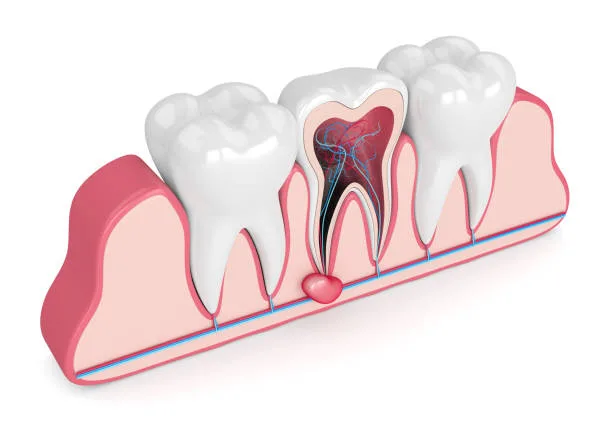
Dental Cyst Treatment: Causes of Formation, Symptoms, and Surgical Procedures
The oral and maxillofacial region, by virtue of its anatomical structure, is home to one of the most complex networks of nerves, vessels, and bones in the body. The healthy attachment of teeth to the jawbone depends on the protection of the living tissue inside them (pulp) and the surrounding supporting tissues (periodontium) from infections. However, as a result of trauma, deep decay, or developmental factors, pathological formations can occur within the jawbone. Dental cysts are among the leading such pathological formations.
A dental cyst is, in medical terms, a pathological cavity filled with fluid, semi-fluid, or gas and surrounded by a membrane-like tissue called epithelium. These structures, which grow slowly and insidiously within the jawbone, can, if not detected in time and cleared by medical intervention, resorb the surrounding healthy bone tissue and cause serious anatomical loss. In this article, the processes of dental cyst treatment, the mechanisms by which cysts form, diagnostic methods, and surgical stages are addressed objectively within a scientific framework.
Why Do Dental Cysts Form? What Are the Types of Cysts?
The causes of cysts developing in the jawbone and tooth roots vary according to the medical type of the cyst. In the general literature, jaw cysts are divided into two main groups:
1. Inflammatory Cysts
This is the type of cyst most commonly encountered in clinics. In medicine, it is known as a "Radicular Cyst" (root-end cyst). It usually begins as a result of an untreated deep cavity or physical trauma to the tooth. The nerve and vessel bundle inside the tooth (pulp) loses its vitality (necrosis). The resulting infection spreads from the root tip to the jawbone. The body's immune system surrounds the inflamed area with a membrane in order to prevent the infection from spreading further into the bone. As a result of this defense mechanism, a fluid-filled cyst sac forms.
2. Developmental Cysts
These occur when tissue remnants remain within the jawbone during the embryological formation of the tooth and later transform into a cystic structure. The best-known example is the form called a "Dentigerous Cyst." These cysts usually develop around unerupted wisdom teeth or canine teeth that remain impacted within the jawbone. Although they are not infection-related, as they grow they put pressure on neighboring teeth and cause structural damage.
What Are the Symptoms of a Dental Cyst?
One of the most dangerous medical characteristics of cysts is that, in their initial and development stages, they are mostly "asymptomatic," meaning they cause no symptoms. Because they grow very slowly within the jawbone, they may go unnoticed by patients for a long time. However, as the cyst increases in size or becomes infected (inflamed), it may present with the following symptoms:
Localized Swelling: Hardness or bulging that can be felt from the outside in the gum area corresponding to the root tip where the cyst is located, or on the jawbone.
Tooth Discoloration: The tooth causing the cyst may take on a gray, brown, or purplish color over time because it has lost its vitality.
Pain and Sensitivity: When the cyst leaves its sterile form and turns into an acute infection, severe, throbbing pain begins in that area. Sensitivity increases when pressure is applied.
Tooth Loosening (Mobility) and Displacement: As the cyst grows, it erodes the jawbone. Tooth loosening may be seen in teeth that have lost their bone support, or shifts in tooth alignment may occur as a result of mechanical pressure exerted on the roots of neighboring teeth.
Numbness (Paresthesia): Massive cysts growing in the lower jaw may create a temporary numbness in the lip and chin when they press on the inferior alveolar nerve.
Diagnosis and Radiological Imaging
Definitive diagnosis of dental cysts cannot be made by visual clinical examination alone. Radiological imaging is essential. A standard panoramic X-ray clearly shows the dark (radiolucent) cyst cavity in the jawbone.
In current clinical diagnostic protocols, Dental Volumetric Tomography (DVT) is used to assess the cyst's exact boundaries, three-dimensional volume, and millimetric relationship with surrounding anatomical structures (sinus cavities, nerve canals). The tomography data determine the roadmap for the surgical treatment to be applied.
Dental Cyst Treatment: Clinical and Surgical Methods
Depending on the size of the cyst, its location, and the condition of the associated tooth, different treatment protocols are applied by physicians. The medical principle is, first and foremost, to completely remove the cyst from the jawbone and, if possible, to keep the tooth that caused the cyst in the mouth.
1. Endodontic Treatment (Root Canal Treatment) and Follow-up
In root-end lesions detected at an early stage and of very small size, sometimes performing only a flawless root canal treatment may be sufficient without the need for surgery. When the infection source inside the tooth is cleaned and the root canals are filled in a sealed manner, the immune system may heal the small cystic structure at the root tip on its own over time. This is monitored by the physician through 6-month radiological follow-ups.
2. Apical Resection (Root-End Surgery)
This is a surgical procedure applied in cysts that do not respond to root canal treatment or are too large to improve with root canal treatment. Under local anesthesia, the gum where the cyst is located is opened with a small incision. The cyst sac in the jawbone is scraped out (enucleation) and cleaned in one piece together with its surrounding membrane. To increase the success of the procedure, approximately 3 millimeters of the root tip of the relevant tooth are cut and removed, and the exposed part of the root tip is sealed from the back (retrograde) with special biocompatible filling materials. The gum is then sutured closed.
3. Tooth Extraction and Cyst Removal
If the cyst has destroyed the tooth root and surrounding bone beyond salvage, or if the relevant tooth is an impacted wisdom tooth, tooth extraction is decided as a medical necessity. After the tooth is extracted, the cyst tissue inside the jawbone is completely cleaned through the extraction socket (curettage). Depending on the size of the cleaned cavity, the physician may place bone graft material in this area.
4. Marsupialization (Cyst Reduction Technique)
This is a staging technique used in cysts that have reached very large sizes and, if removed in a single procedure, carry a risk of breaking the jawbone or harming important nerves. A small window is opened over the cyst, the fluid inside is drained, and the pressure inside the cyst is reduced. As the pressure decreases, the cyst gradually shrinks away from the bone over the course of months. When the cyst has been surgically reduced to a safe size, it is completely removed in a second operation.
Recovery Process and Care After Surgery
Because dental cyst operations are mostly performed under local anesthesia, the patient does not feel pain during the procedure. However, the post-operative period is extremely critical for successful healing:
Cold Compress: During the first 24 hours, applying ice externally to the cheek at intervals specified by the physician (cold compress) minimizes edema (swelling) and bruising that may occur.
Medical Support: To prevent the risk of infection after the operation, the antibiotics, painkillers, and special mouth rinses prescribed by the physician must be used without interruption and at the full dose.
Nutrition and Hygiene: For the first few days, chewing should not be done on the operated area, and warm, soft foods should be consumed. Tobacco products must absolutely not be used for the first 72 hours, as they will disrupt clotting and stop wound healing. The sutured area should be kept clean gently, without brushing against it.
Risks Posed by Untreated Cysts
Dental cysts are not structures that can be neglected with the thought that they will "go away over time." A cyst left untreated in the bone continues to grow over the years. As a result of this growth, the cortical (hard) layer of the jawbone thins, and jawbone fractures (pathological fractures) can occur even with a simple trauma. In addition, the spread of infection into the bloodstream (focal infection) and the cyst eroding the roots of neighboring teeth, causing those teeth to be lost as well, are possible medical scenarios.
All removed pathological tissues are sent to the pathology laboratory, as a standard medical procedure, for examination of their cellular structure. This is a routine step taken to establish the definitive diagnosis of the tissue.
For a healthy jaw structure, it is very important that you do not neglect your routine dental checkups and that you consult a specialist clinic without delay if you notice any abnormal swelling in the mouth.
Legal Notice: This article has been prepared in line with the regulations on Promotional and Informational Activities in Healthcare Services, with the aim of informing the public about health and raising awareness. The statements in the text can in no way replace a professional examination, clinical diagnosis, or personalized treatment planning by a specialized physician or dentist. Each patient's anatomical structure and treatment process are different. For definitive diagnosis and medical guidance regarding dental diseases, maxillofacial surgery, and all other clinical procedures, it is imperative to consult an authorized oral and dental health institution.